
19 UMN Students Join International Student Exchange Program
August 24, 2026
Why Should College Students Start Learning Data Analytics Early?
August 20, 2026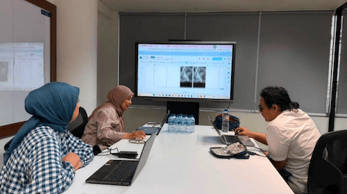
Discussion to assess the accuracy of AI and Augmented Reality in detecting kidney lesions (back left): Monica; (front left): Irmawati; (right): Dr. Edvin. Photo: personal collection
Jakarta (August 13, 2026) – A research team from Multimedia Nusantara University (UMN), in collaboration with the Faculty of Medicine at Brawijaya University, is working to develop Artificial Intelligence (AI) and Augmented Reality to assist doctors in detecting kidney abnormalities.
The UMN research team consists of Dr. Irmawati, S.Kom., M.M.S.I., a lecturer in the Faculty of Information Technology, and Monica Pratiwi, S.ST., M.T., a lecturer in the Faculty of Computer Engineering. Meanwhile, the clinical team consists of Dr. Edvin Prawira Negara, Dr. Alfryan Janardhana, Dr. Ahmad Irsyad Syafrani, and Dr. Besut Daryanto, Sp.B., Sp.U(K), from the Department of Urology. The research also involved Prof. Dr. dr. Yuyun Yueniwati, Sp.Rad(K), from the Department of Radiology at the Regional General Hospital, Dr. Saiful Anwar, and the Faculty of Medicine at UB.
“Collaboration between technology researchers and doctors is crucial to ensure that the AI developed truly addresses healthcare needs and can be used appropriately in clinical settings,” said Dr. Irmawati from UMN in her press release.
Through this research, the joint team aims to help detect kidney abnormalities—such as kidney stones and kidney cancer—using AI and Augmented Reality (AR) technology. The technology utilizes CT scan images and employs radiomic analysis methods. This method converts medical images into quantifiable data. The hope is that the resulting data will provide information regarding the shape, size, texture, location, and characteristics of tissues that are not always easily identifiable through visual observation.
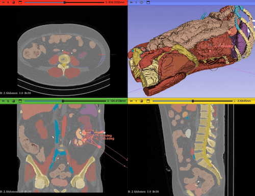
Automatic lesion detection and surgical simulation performed on a patient
The AI then processes this data and uses Augmented Reality to display the analysis results in a three-dimensional visual format. This is expected to help doctors better visualize the position of the kidneys, the location of stones or tumors, and their relationship to surrounding blood vessels and tissues.
This technological system is expected to help doctors make more accurate diagnoses, plan treatments, and prepare for surgeries more effectively. Furthermore, the visual representations generated by Augmented Reality are expected to make it easier for doctors to explain a patient’s condition and treatment plan to their family.
So far, this research has successfully developed a kidney radiomics AI model, a feasibility study report, and a web-based application to process and display medical image analysis results. The next step is to conduct further testing and clinical validation before this technology can be widely used to serve patients.
By Putu Wiena | UMN News Service
English translation by Levina Chrestella Theodora
Kuliah di Jakarta untuk jurusan program studi Informatika| Sistem Informasi | Teknik Komputer | Teknik Elektro | Teknik Fisika | Akuntansi | Manajemen| Komunikasi Strategis | Jurnalistik | Desain Komunikasi Visual | Film dan Animasi | Arsitektur | D3 Perhotelan , di Universitas Multimedia Nusantara.